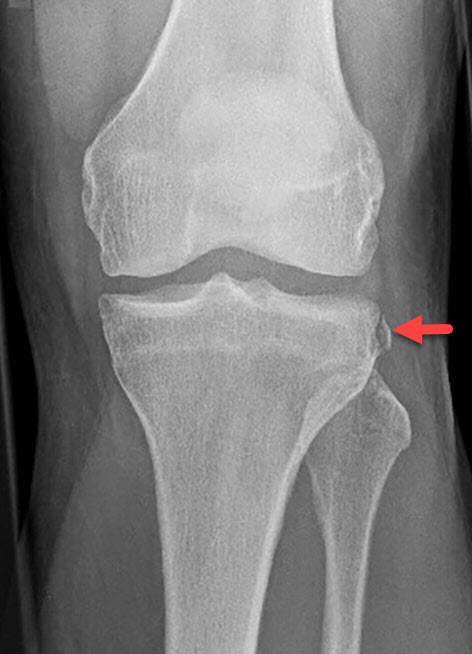
A Segond Fracture is a small avulsion fracture involving the lateral aspect of the knee just above the fibula head. In approximately 75% of the cases, it is the first indication of an anterior cruciate ligament tear. you should recommend MRI scan as a follow-up whenever you see the type of fracture.

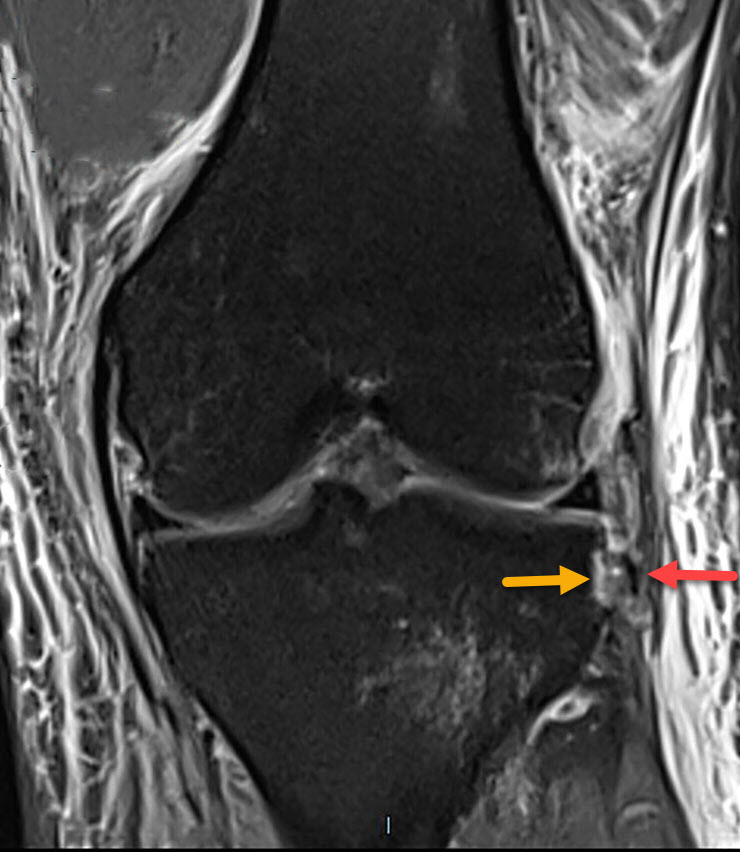
The red arrow points at the small avulsion fracture and the yellow arrow points at a defect caused by the avulsion fracture at the lateral aspect of the tibia. There is a lot of soft tissue edema on both the medial and lateral aspects of the knee. A little bit of edema in the lateral proximal tibia

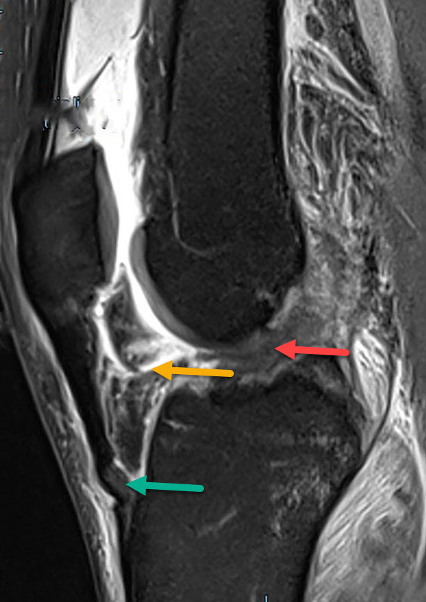
Full-thickness Anterior Cruciate Ligament(ACL) tear in the same patient on a follow-up MRI scan. Red arrow points at the full-thickness ACL tear. Yellow arrow points at a laceration in the Hoffa fat pad and the green arrow points at a tendinopathy of the patella ligament. There is a lot of soft-tissue edema and hemarthrosis in the knee joint.

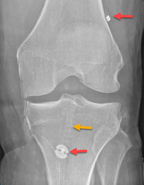
The X-Ray diagram above shows the graft used to replace the ACL tear in the same patient. The yellow arrow points at a tunnel through which the graft travels. The red buttons point at the attachment sites. Note: the Segond Fracture can still be seen from the original fracture on the lateral aspect of the proximal tibia above the fibula head.
